sCT Generation from Nuclear Medicine Scans
A Patch-Based Ensemble Approach to Generate High-Fidelity Synthetic CT (sCT) Images from PET Scans
The Challenge: Adding Anatomy to Function
Nuclear Medicine Scan (PET)
Reveals metabolic function but lacks detailed anatomical information.
High-Fidelity Synthetic CT
Provides rich anatomical context, enhancing diagnostic value and treatment planning.
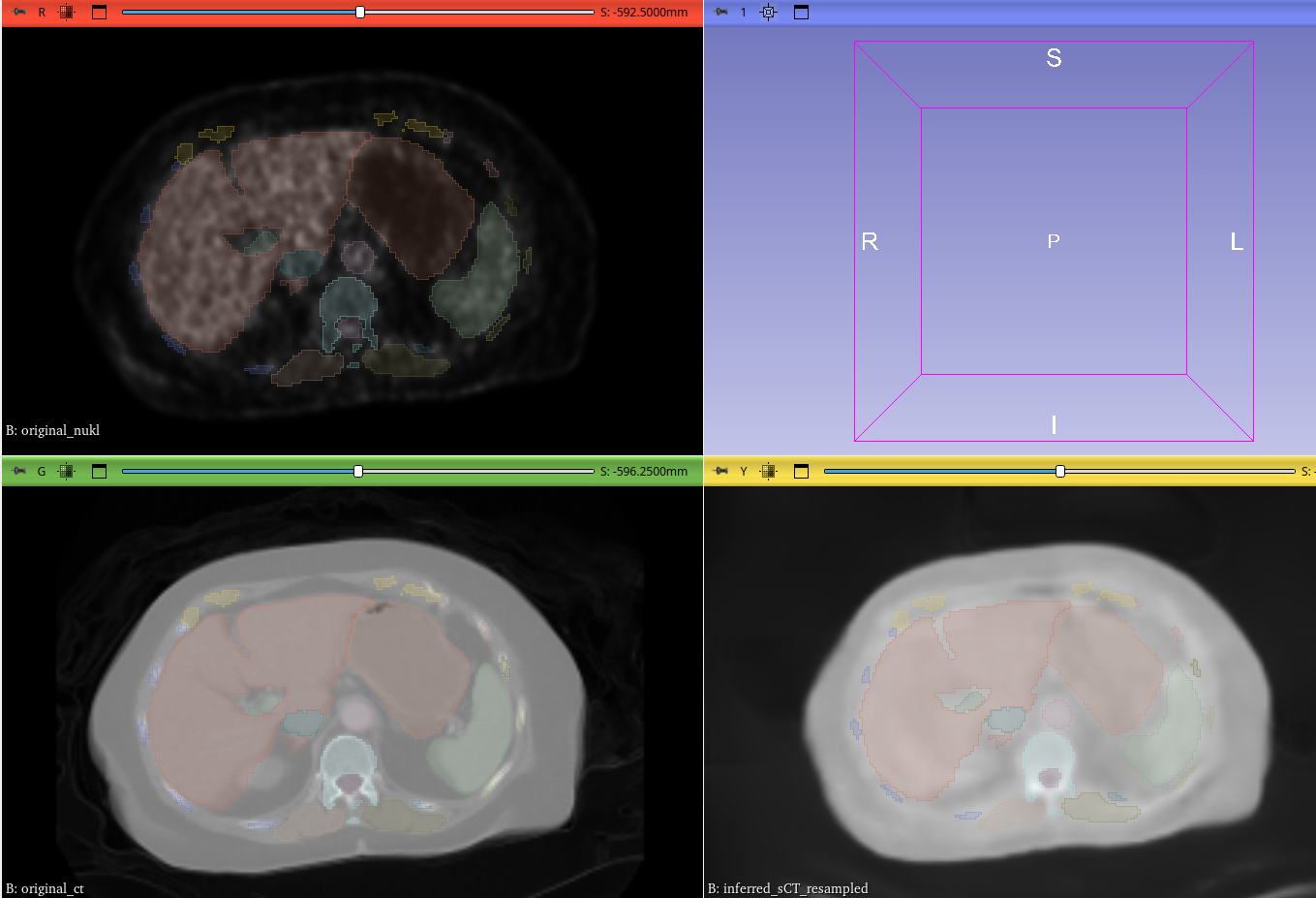
Visualizing the Results
PET vs. Original CT vs. Synthetic CT
This comparison highlights the anatomical accuracy of our synthetic CT (right), which closely mirrors the ground truth of the original CT (middle) while retaining the functional context from the PET scan (left).
Key Innovations & Expertise
1. The Ensemble of Experts
Five independent nnU-Net models, pretrained as "experts" on specific anatomical regions, work together to generate a highly detailed sCT.
2. The “Anatomical Supervisor”
A frozen, parallel ensemble of models acts as a "supervisor," constantly evaluating the generated sCT for anatomical correctness and sending a strong loss signal to correct errors.
3. Efficient Patch-Based Training
The model trains on smaller 128x128x128 patches of the full-resolution image, overcoming memory limitations and making advanced model development feasible.
Clinical Impact & Significance
Enhance Diagnostic Confidence
Provides crucial anatomical context for functional scans.
Improve Attenuation Correction
Leads to more accurate quantification in PET imaging.
Enable More Precise Treatment Planning
Allows clinicians to better localize tumors and pathologies.